Quantitative Membrane Biophysics
The ongoing research topics are given below:
The Physics of Morphological Instabilities.
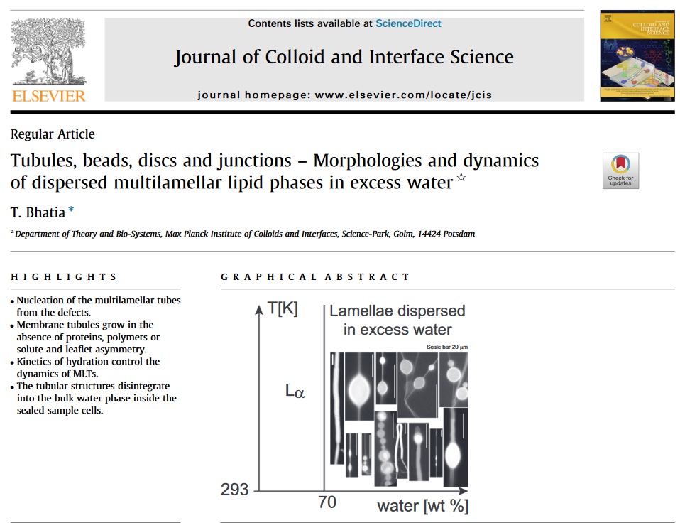
The Physics of Biomembrane Dynamics.
Thermodynamics of Biological Membranes.
Simulations & AI.
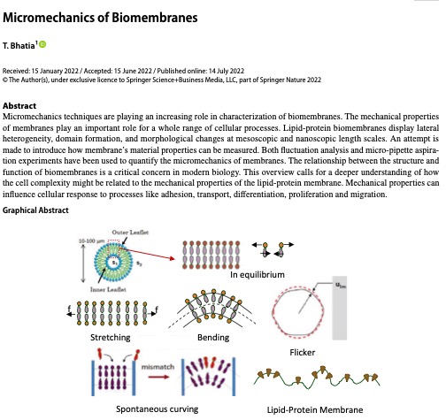
Micromechanics of Biological Membranes.
Light and Matter Physics.
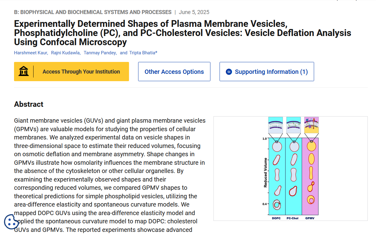
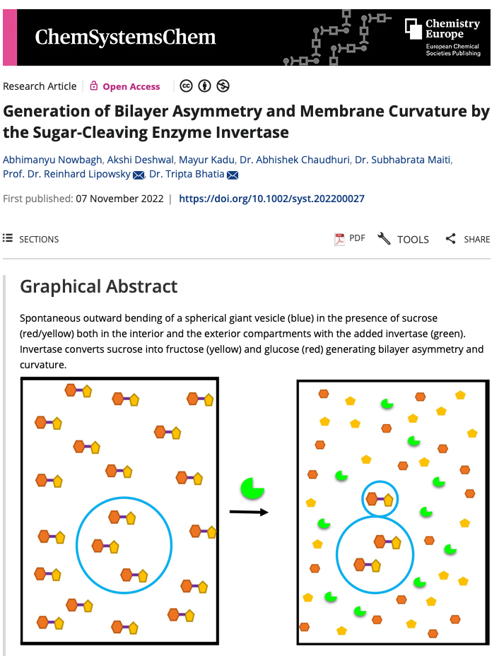
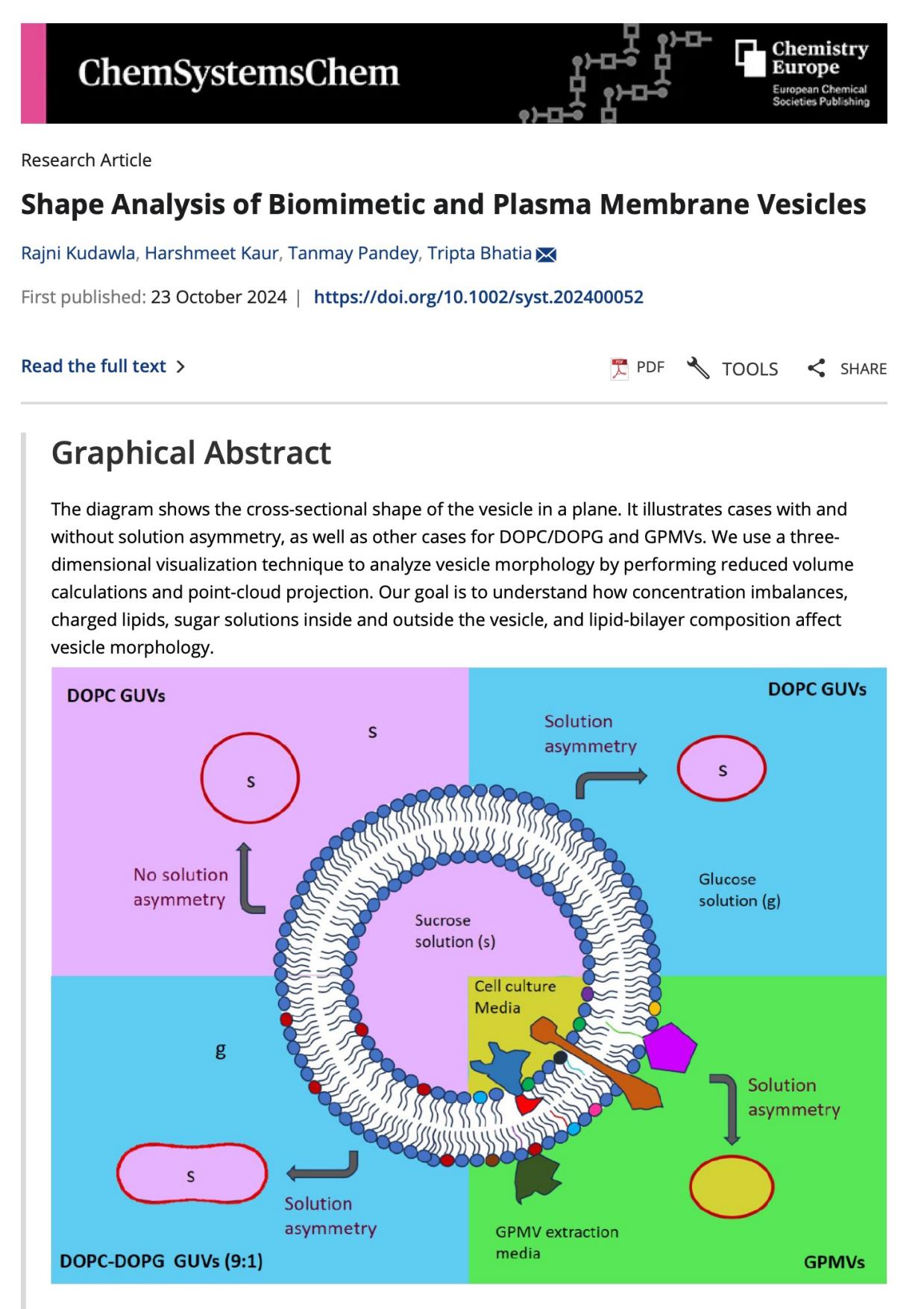
Biophysics of Cell-derived Vesicles.
[Time-bound projects are available with defined start and end dates.]
 IRINS
IRINS